• Request Info
  • Visit
  • Apply
  • Give
  • Request Info
  • Visit
  • Apply
  • Give

Search

  • A-Z Index
  • Map

Microbiology

  • About
    • Alumni and Friends
    • Annual Awards Day Ceremony
    • Facilities Resources
    • News
    • REU Program
  • People
    • Faculty
    • Affiliate Faculty
    • Staff
    • Research and Training Faculty
    • Emeritus
    • Post Doctoral
    • Current Graduate Students
    • Graduate Student Alumni
  • Research
  • Undergraduate
    • Advising
    • Courses
    • Course Offerings By Semester
    • FAQs
    • Honors in Microbiology Guidelines
    • MUC
  • Graduate
    • Assistantships
    • Course Descriptions
    • Steps to Graduation
    • Frequently Asked Questions
  • Seminars & Events
    • Seminar Series
    • Student Colloquium

The Conversation: ‘Mitochondria can sense bacteria and trigger your immune system to trap them – revealing new ways to treat infections and autoimmunity’

June 30, 2025 by Logan Judy

no photo placeholder image

The Conversation: ‘Mitochondria can sense bacteria and trigger your immune system to trap them – revealing new ways to treat infections and autoimmunity’

Profile
Neutrophils (yellow) eject a NET (green) to ensnare bacteria (purple). Other cells, such as red blood cells (orange), may also get trapped. CHDENK/Wikimedia Commons, CC BY-SA
Andrew Monteith, University of Tennessee

Mitochondria have primarily been known as the energy-producing components of cells. But scientists are increasingly discovering that these small organelles do much more than just power cells. They are also involved in immune functions such as controlling inflammation, regulating cell death and responding to infections.

Research from my colleagues and I revealed that mitochondria play another key role in your immune response: sensing bacterial activity and helping neutrophils, a type of white blood cell, trap and kill them.

For the past 16 years, my research has focused on understanding the decisions immune cells make during infection and how the breakdown of these decision-making processes cause disease. My lab’s recent findings shed light on why people with autoimmune diseases such as lupus may struggle to fight infections, revealing a potential link between dysfunctional mitochondria and weakened immune defenses.

Side-by-side comparison of labeled illustration of cross-section of mitochondria and its micrograph
Mitochondria do so much more than just produce energy. OpenStax, CC BY-SA

The immune system’s secret weapons

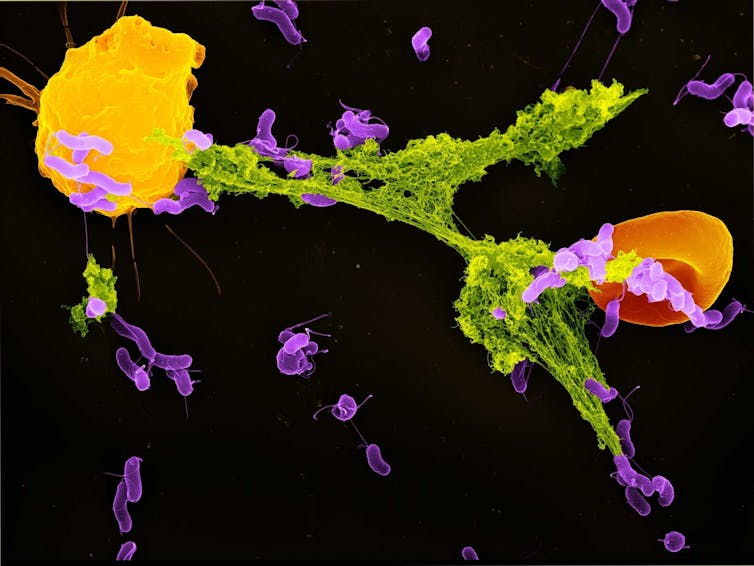
Neutrophils are the most abundant type of immune cell and serve as the immune system’s first responders. One of their key defense mechanisms is releasing neutrophil extracellular traps, or NETs – weblike structures composed of DNA and antimicrobial proteins. These sticky NETs trap and neutralize invading microbes, preventing their spread in the body.

Until recently, scientists believed that NET formation was primarily triggered by cellular stress and damage. However, our study found that mitochondria can detect a specific bacterial byproduct – lactate – and use that signal to initiate NET formation.

Lactate is commonly associated with muscle fatigue in people. But in the context of bacterial infections, it plays a different role. Many bacteria release lactate as part of their own energy production. My team found that once bacteria are engulfed by a compartment of the cell called the phagosome, neutrophils can sense the presence of this lactate.

Inside the phagosome, this lactate communicates to the neutrophil that bacteria are present and that the antibacterial processes are not sufficient to kill these pathogens. When the mitochondria in neutrophil cells detect this lactate, they start signaling for the cell to get rid of the NETs that have entrapped bacteria. Once the bacteria are released outside the cell, other immune cells can kill them.

Here, a neutrophil engulfs MRSA bacteria (green).

When we blocked the mitochondria’s ability to sense lactate, neutrophils failed to produce NETs effectively. This meant bacteria were more likely to escape capture and proliferate, showing how crucial this mechanism is to immune defense. This process highlights an intricate dialogue between the bacteria’s metabolism and the host cell’s energy machinery.

What makes this finding surprising is that the mitochondria within cells are able to detect bacteria trapped in phagosomes, even though the microbes are enclosed in a separate space. Somehow, mitochondrial sensors can pick up cues from within these compartments – an impressive feat of cellular coordination.

Targeting mitochondria to fight infections

Our study is part of a growing field called immunometabolism, which explores how metabolism and immune function are deeply intertwined. Rather than viewing cellular metabolism as strictly a means to generate energy, researchers are now recognizing it as a central driver of immune decisions.

Mitochondria sit at the heart of this interaction. Their ability to sense, respond to and even shape the metabolic environment of a cell gives them a critical role in determining how and when immune responses are deployed.

For example, our findings provide a key reason why patients with a chronic autoimmune disease called systemic lupus erythematosus often suffer from recurrent infections. Mitochondria in the neutrophils of lupus patients fail to sense bacterial lactate properly. As a result, NET production was significantly reduced. This mitochondrial dysfunction could explain why lupus patients are more vulnerable to bacterial infections – even though their immune systems are constantly activated due to the disease.

This observation points to mitochondria’s central role in balancing immune responses. It connects two seemingly unrelated issues: immune overactivity, as seen in lupus, and immune weakness like increased susceptibility to infection. When mitochondria work correctly, they help neutrophils mount an effective, targeted attack on bacteria. But when mitochondria are impaired, this system breaks down.

Microscopy image of long threads extending from round blobs
Neutrophils unable to effectively produce NETs may contribute to the development of lupus. Luz Blanco/National Institute of Arthritis and Musculoskeletal and Skin Diseases via Flickr, CC BY-NC-SA

Our discovery that mitochondria can sense bacterial lactate to trigger NET formation opens up new possibilities for treating infections. For instance, drugs that enhance mitochondrial sensing could boost NET production in people with weakened immune systems. On the flip side, for conditions where NETs contribute to tissue damage – such as in severe COVID-19 or autoimmune diseases – it might be beneficial to limit this response.

Additionally, our study raises the question of whether other immune cells use similar mechanisms to sense microbial metabolites, and whether other bacterial byproducts might serve as immune signals. Understanding these pathways in more detail could lead to new treatments that modulate immune responses more precisely, reducing collateral damage while preserving antimicrobial defenses.

Mitochondria are not just the powerhouses of the cell – they are the immune system’s watchtowers, alert to even the faintest metabolic signals of bacterial invaders. As researchers’ understanding of their roles expands, so too does our appreciation for the complexity – and adaptability – of our cellular defenses.The Conversation

Andrew Monteith, Assistant Professor of Microbiology, University of Tennessee

This article is republished from The Conversation under a Creative Commons license. Read the original article.

Filed Under: Faculty, Featured, News

Alison Buchan named 2024 AAAS Fellow

April 9, 2025 by Logan Judy

no photo placeholder image

Alison Buchan named 2024 AAAS Fellow

Profile

Filed Under: Faculty, Featured, News

Monteith Lab Tracks Immune Response

February 27, 2025 by Logan Judy

no photo placeholder image

Monteith Lab Tracks Immune Response

Profile

Filed Under: Faculty, Featured, News

Mikucki Digs Deep for Space-Bound Microbiology

January 15, 2025 by Logan Judy

no photo placeholder image

Mikucki Digs Deep for Space-Bound Microbiology

Profile

Filed Under: Faculty, Featured, News

Research Takes UT Faculty and Students to the Extreme

December 10, 2024 by Logan Judy

no photo placeholder image

Research Takes UT Faculty and Students to the Extreme

Profile

Filed Under: Faculty, Featured, News

$1.82 million NIH Grant Funding Lindsey Burcham’s Women’s Health Research

July 24, 2024 by Logan Judy

no photo placeholder image

$1.82 million NIH Grant Funding Lindsey Burcham’s Women’s Health Research

Profile

by Amy Beth Miller

With a $1.82 million National Institutes of Health grant, Assistant Professor Lindsey Burcham is leading interdisciplinary research on the vaginal microbiome, which can affect maternal, fetal, and pediatric health.

“We are incorporating techniques in microbial genetics/molecular biology, analytical chemistry, and in vitro and in vivo models to learn more about how microbes persist in the vaginal mucosa,” Burcham explained.

“This work will allow us to learn more about the availability of micronutrients in the vaginal tract and understand how vaginal microbes sense nutrient availability, respond to nutrient fluctuations, and share these nutrients within the community,” she said.

The five-year Maximizing Investigators’ Research Award (MIRA) began in 2023 and will run until June 2028. 

Cultures and Computer Models

Burcham also is using computer simulations, or in silico models, in collaboration with UT microbiology Assistant Professor Zach Burcham to predict metabolite exchange between vaginal microbes.

“These models allow us to develop testable hypotheses to understand more about how microbes may interact with each other and behave in the host,” Lindsey Burcham said.

The researchers also will use synthetic communities, or controlled co-cultures of microbes, to study microbial interactions and to understand how individual microbes may impact the overall function of the microbial community.

Foundations for Exploration 

The MIRA funding provides flexibility for investigators to be creative and work toward big-picture questions, Lindsey Burcham said. “The work outlined here will set the foundation for understanding more about the vaginal environment and microbe-microbe interactions, but I envision this is a starting point. We have already started some exciting new projects investigating other environmental factors within the vaginal tract, and we will go where the data take us.”

She has helped to assemble a collaborative “bench-to-bedside” research team including Zach Burcham and physicians Kim Fortner, Callie Reeder, and Logan Riley, as well as Associate Professor Jill Maples from the University of Tennessee Medical Center’s Department of Obstetrics and Gynecology.

Lindsey Burcham has been curious about microbes and motivated to understand how they work since she was an undergraduate student, and her personal life influenced her research interests. 

“My own pregnancies fueled my curiosity for understanding the vaginal tract and the impact of microbes on vaginal health,” she said. “Now as the leader of a research team, I am excited to be able to use my lab and resources to promote women’s health research and to normalize the discussion around vaginal health.”

Filed Under: Faculty, Featured

UT Faculty Join New $6.5 Million Grant to Study Climate Links Between Algal Blooms and Human Health

June 3, 2024 by Logan Judy

no photo placeholder image

UT Faculty Join New $6.5 Million Grant to Study Climate Links Between Algal Blooms and Human Health

Profile

Faculty in the University of Tennessee Department of Microbiology are part of a new $6.5 million, five-year federal grant to establish a virtual center for the study of links between climate change, harmful algal blooms, and human health.

Increased precipitation, more powerful storms, and warming Great Lakes waters all encourage the proliferation of harmful algal blooms composed of cyanobacteria, also known as blue-green algae. These microorganisms can produce toxins harmful to humans, pets, and wildlife.

Though the pea-green summer blooms in western Lake Erie are the best-known in the region, cyanobacterial harmful algal blooms, or cHABs, now occur in all five Great Lakes and in fresh waters around the US. These blooms can increase water-treatment costs for local governments and harm vital summer economies in communities that enjoying fishing, swimming, and boating. In the Knoxville area earlier this year, a cHAB of Planktothrix rubescens (“pink algae”) in Meads Quarry at Ijams Nature Center closed swimming and other activity for a month.

Student researchers collect water samples from a cyanobacterial harmful algal bloom in western Lake Erie as part of a project to study the effect of environmental conditions on toxin production by cyanobacteria. Photo by McKenzie Powers

“The University of Tennessee has a 25-plus year history of contributing to research on the security of our water supplies with respect to harmful algal blooms, with the Great Lakes being a major focus,” noted Steven Wilhelm, the Kenneth and Blaire Mossman Professor of Microbiology and one of the project’s leaders.

The UT team, including Wilhelm and David Talmy, assistant professor in the Department of Microbiology, will focus on the effects of a changing climate and identify new players in the biological problems the lake is having. To understand the drivers of toxic algal bloom events, they previously collaborated with researchers in Michigan and Berlin, Germany, on the development of mathematical models that predict toxin production by cells, and how some of the proposed abatement methods may have unintended consequences.

Understanding best abatement practices and projecting the long-term effects of a changing climate—as well as the disruptive storms that go with it—are a major goal of this research. Participants will work across four collective projects to enable an assessment of the human health risks of cHAB toxins. These complementary projects seek to determine how climate change affects cHABs and how cHABs impact human health.

This virtual center was founded at Bowling Green State University in 2018 with funding from the National Institutes of Health and the National Science Foundation. Following the retirement of founding director Professor George Bullerjahn at BGSU, the center’s administrative home moved to Ann Arbor with renewed funding from the two federal agencies under the directorship of University of Michigan (UM) Professor Gregory Dick.

The center maintains core projects as it evolves to pursue new directions informed by more than 70 research papers published in peer-reviewed scientific journals by center-funded scientists since 2018. In addition, center-funded researchers will develop new technologies for advanced monitoring and forecasting of cyanobacterial harmful algal blooms in collaboration with colleagues at NOAA’s Great Lakes Environmental Research laboratory and the U-M-based Cooperative Institute for Great Lakes Research.

“The threat to water resources in the Great Lakes—which hold about 95% of the surface fresh water in the US and support a multibillion-dollar blue economy—is real,” said Dick. “But despite these serious threats, key scientific questions surrounding the climate drivers and health impacts of cHABs remain unresolved. Our knowledge is not yet sufficient to predict how a changing climate will impact cHAB distributions, community composition, or toxicity.” 

Studies will combine observation, experiment, and modeling in areas of lake science, climatology, microbiology, and biomedical science. A community engagement core led by The Ohio State University (OSU) will communicate findings to relevant communities and stakeholders.

In addition to UT, UM and OSU, partners include more than 28 faculty researchers and dozens of students at Bowling Green State University, the University of Toledo, Wayne State University, Michigan State University, University of North Carolina, James Madison University, the State University of New York, and the University of Windsor in Canada.

By Randall Brown

Filed Under: Faculty, Featured, News

Zepernick Investigates How Freshwater Diatoms Stay in the Light

March 13, 2024 by Logan Judy

no photo placeholder image

Zepernick Investigates How Freshwater Diatoms Stay in the Light

Profile

Spring weather brings welcome conditions for flowers and plant life to bloom across the land. The right mixture of temperature, moisture, and light helps keep the green world vibrant.

Underwater plant life generally responds to similar environmental encouragements, but a curious discovery in Lake Erie circa 2012 led microbiologists to study an unseasonal display of winter abundance. Blooms of diatoms—microscopic, photosynthetic algae—were alive and well beneath (and within) the lake’s ice cover.

“Some of the main winter-spring diatom bloom formers, like Aulacoseira islandica, have a symbiotic relationship with heterotrophic bacteria capable of forming tiny ice crystals, which over time causes the diatom filaments to become buoyant—just as ice cubes float in your favorite beverage,” said Brittany Zepernick, a post-doctoral researcher and SEC Emerging Scholar in UT’s Department of Microbiology.

These ‘diatom ice cubes’ float to the Lake Erie ice cover and embed within it, putting them in position to absorb the light needed to perform photosynthesis throughout the winter months. It was good news for diatoms, which are a vital component of the cumulative ecosystem in lakes and oceans across the globe

This curious adaptation is threatened, though, as warming global temperatures have led to widespread ice decline across the Great Lakes, leaving Lake Erie in a nearly ice-free state in several recent winters and leaving diatoms stuck in murky, light-deprived waters. In these new “climatically uncharted waters,” the adaptations that benefitted these winter diatoms for so long suddenly ceased to serve them.

So, what’s a diatom to do? Zepernick and colleagues turned to the shores of Lake Erie to investigate the evolving situation. With the help of the US and Canadian Coast Guard, they sampled the ice-covered (in 2019) and ice-free (in 2020) winter waters of Lake Erie to learn how diatoms were responding to changing environmental conditions. They recently published their work in the ISME Journal—Multidisciplinary Journal of Microbial Ecology.

Two main diatom genera dominate the winter blooms: Aulacoseira islandica and Stephanodiscus spp.

“The abundance of Stephanodiscus spp. was approximately 70 percent lower in the ice-free water column of 2020 compared to the ice-covered water column of 2019,” said Zepernick. “Likewise, the abundance of Aulacoseira islandica was around 50 percent lower in the ice-free water column compared to the ice-covered water column.”

Ice floating on the surface of Lake Erie
Diatoms adapted to embed in Lake Erie’s ice cover—can they adapt to stay afloat without ice?

With ice cover across the Great Lakes at record lows—from around 80 percent covered in ice in 2018 and 2019 to just 8 percent covered in 2023—researchers expect this trend will continue in future winters. 

The next step is studying how this impacts Lake Erie, which joins the other Laurentian Great Lakes of the US and Canada to cumulatively contain approximately 20 percent of the globe’s fresh water.

“Despite the critical importance of this system, we didn’t know diatom blooms even formed in the winter-spring months until around 2012,” said Zepernick. “Many researchers have referred to the winter water column as a ‘New Frontier’ or a ‘black box.’ What we do know is that diatoms are critically important to regional lake ecosystems and global climate.”

Diatoms make up an estimated 20 percent of global carbon sequestration and oxygen production, play an enhanced role in global biogeochemical cycles, and represent a critical component of the aquatic ecosystem in freshwater systems.

“Hence, the large-scale changes already underway to the winter-spring diatom communities in Lake Erie and other lakes across the globe will result in large-scale biological and biogeochemical change,” said Zepernick.

The light at the end of the icy tunnel could rely on the diatoms’ potential to adapt. Zepernick’s recent work indicates they could possibly form clusters with adhesive proteins called fasciclins to “raft” to the surface of the muddy waters via “underwater waves” produced by wind, convection, and underwater currents.

Another adaptation Zepernick hinted at was that diatoms could increase their use of proton-pumping rhodopins (PPRs)—light harvesting, retinal-containing proteins that could serve as an alternative to classical photosynthesis. She is currently attempting to isolate freshwater diatoms from Lake Erie samples that possess PPRs to create a model freshwater diatom-PPR system for further study. Her findings could offer clues to the diatoms’ next move in a rapidly changing climate.

“PPRs are a hot topic within marine literature, yet we know very little about how these mechanisms apply to freshwater systems and taxa,” she said. “I am interested in elucidating the benefits PPRs may confer to both freshwater and marine diatoms across a variety of emerging—and future—climatic stressors.”

Filed Under: Faculty, News

Recent Posts

  • Scholar Spotlight: Zachary Burcham
  • We’re Hiring!
  • UT’s Rising Stars of Microbiology
  • The Conversation: ‘Mitochondria can sense bacteria and trigger your immune system to trap them – revealing new ways to treat infections and autoimmunity’
  • Alison Buchan named 2024 AAAS Fellow

Recent Comments

No comments to show.

College of Arts & Sciences

117 Natalie L. Haslam Music Center
1741 Volunteer Blvd.
Knoxville TN 37996-2600

Phone: 865-974-3241

Archives

  • September 2025
  • August 2025
  • June 2025
  • April 2025
  • February 2025
  • January 2025
  • December 2024
  • August 2024
  • July 2024
  • June 2024
  • April 2024
  • March 2024
  • September 2023
  • August 2023
  • May 2023
  • April 2023
  • March 2023
  • February 2023
  • January 2023
  • July 2022
  • January 2020

Categories

  • Faculty
  • Featured
  • News
  • Newsletter
  • Uncategorized

Copyright © 2025 · University of Tennessee, Knoxville WDS Genesis Child on Genesis Framework · WordPress · Log in

Department of Microbiology

College of Arts and Sciences

1311 Cumberland Avenue
307 Ken and Blaire Mossman Bldg.
Knoxville TN 37996-1937

Email: microbiology@utk.edu

Phone: 865-974-3441

The University of Tennessee, Knoxville
Knoxville, Tennessee 37996
865-974-1000

The flagship campus of the University of Tennessee System and partner in the Tennessee Transfer Pathway.

ADA Privacy Safety Title IX