Looking closely at microplastics
by Hanna Boshnag
Microplastics seems to be a big buzzword these days. It’s easy to roll one’s eyes at another sensational news headline, but these could have truly harmful impacts on the environment. The use of plastic mulching in agriculture could alter the soil microbiome and have adverse effects on the broader ecosystem. Biodegradable mulch is a potential alternative, but it’s uncertain how significant of a difference it makes.
Toyosi Nimat Ajide-Bamigboye, a PhD candidate at the University of Tennessee, Knoxville, is working to characterize this difference.
“Both biodegradable and non-biodegradable mulches generate fragments in the environment, which I study to see how it affects microorganism growth and function,” she said.
To ensure they are not inadvertently releasing microplastics into the environment, the lab collects soil from a field and uses a greenhouse as a controlled environment. After mulch is laid down, Ajide-Bamigboye checks a variety of factors over the course of six months: soil pH, degradation, and electrical conductivity. Once a month, a sample is sent off for sequencing to map changes in the microbiome.
This is a long and tedious process, which Ajide-Bamigboye says she enjoys every second of. She is passionate about every part of the process of doing research because it’s a working step to discovering something new.
Ajide-Bamigboye grew up in Nigeria, where she studied microbiology for both her bachelor’s and master’s degrees. When she decided to pursue her PhD at UT, she brought her husband and two children with her. Despite the difficulties of moving her family, she credits them as the reason she’s able to wholeheartedly work on her research.
“I have a five-year-old daughter and three-year old son,” she said. “My husband is my support system, and when I’m in school, I know I don’t have to worry about them because I know he has it under control.”
After completing her PhD, Ajide-Bamigboye hopes to stay with her family here and work in industry, using her research and skills to contribute creative solutions in the field of agricultural microbiology.
Increasing the Speed of Scientific Discovery
by Hanna Boshnag
Having been involved in conservation in high school, Liz Glasgo was excited to study biology as an undergraduate. When she got to Bowling Green State University, her catalyst for choosing microbiology was learning about marine microbes: diatoms, dinoflagellates, and radiolarians.
“I thought they were so interesting and beautiful to look at,” she said.
Later her undergraduate research focused on freshwater microbiology. After completing her bachelor’s degree, she chose to come to the University of Tennessee because of the sense of community within the Department of Microbiology.
Glasgo is a part of the Zinser Lab, where her research centers on Vibrio natriegens, a fast-growing, Gram-negative marine bacterium. She is attempting to characterize its oxidative stress response and fitness outcomes following genome reduction.
“V. natriegens is an emerging model organism, and the developments of new techniques and a better understanding of its physiology will be helpful to the scientific community,” she said.
For much of microbiology’s history, Escherichia coli has been the primary model organism for researchers to develop new techniques and understand a variety of biological systems. Discoveries as important as the genetic code, DNA replication, gene regulation, and more have been made possible with E. coli as the go-to organism for microbial study.
So why the potential switch to V. natriegens? The difference in growth rate. The marine bacterium has the fastest known generation time and is much quicker to double when compared with E. coli. This shortened growth time introduces the potential of faster experimentation and advanced genomic techniques. Glasgo’s work to better understand V. natriegens genetics and physiology is, thus, all the more important.
To Glasgo, the most exciting aspect of research is directly carrying out her experiments, and being the first to find out something new.
“A lot of my work has involved genetics and making mutants, which entails a lot of PCRs (polymerase chain reactions) and molecular work,” she said. “Day-to-day, you can usually find me in the hood, working with cultures of bacteria and quantifying them on agar plates.”
E. coli might be able to take a break from its Nobel Prize-winning discoveries and make way for V. natriegens, a speedster bacterium with incredible potential for discoveries. Glasgo is particularly excited about its new applications in bioengineering and synthetic biology.
Finding the Balance, in the Lab and Out
by Hanna Boshnag
As a fourth-year PhD candidate, Mikayla Mangrum is well-versed in the art of being a graduate student, both in and out of the lab.
Mangrum’s interest in science and biology began in high school. Microbiology courses during her undergraduate studies at Tennessee Tech University inspired her to study it further. Mangrum decided to pursue her PhD at UT’s Department of Microbiology because of its balance between pathogenesis and ecology—and its proximity to home.
As part of Professor Todd Reynolds’ Microbial Physiology and Pathogenesis Lab, she studies Candida albicans, a common fungus that exists as part of our body’s microbiome. However, when homeostasis is disrupted, it can grow out of control and cause diseases such as thrush and vaginal yeast infections.
The cell wall of C. albicans is made up of many components: mannoproteins, chitin, β-1,6-, and β-1,3-glucan. Together, these can hide it from host immune response and increase resistance to environmental stress. Mangrum’s work focuses on “unmasking” the β-1,3-glucan component of the cell wall to compromise the fungus and, hopefully, discovering ways to target C. albicans infections.
“We try to look at different ways that we can manipulate the cell wall to make it more available to the host immune system and improve host outcome to the fungus,” she explained. “We attempt to do it genetically and with potential drug treatments.”
The concept of unmasking isn’t limited to the study of Candida and could be applied to other fungi as well. In studying the enzymes C. albicans uses to evade the host immune system, Mangrum’s work also could uncover potential fungus-specific drug targets.
To her, two of the most important things a PhD candidate can have in their proverbial toolkit are passion and a strong support system. She explains that passion can be an asset when a scientist encounters setbacks or obstacles in their research, which can be overwhelming. Having a good support system—in your mentor and fellow lab mates—also can make all the difference.
“The PhD is about learning how to learn, rather than being a senior-level professional by the time you leave,” she said. “So having a mentor who is going to support you as a person as well as a scientist is pretty important.”
Outside of her research, Mangrum enjoys hiking, crocheting, and holding game nights with fellow PhD students. She says that having the balance between non-science hobbies and working in science is monumental to maintaining passion and drive for her research.
After completing her doctorate, Mangrum hopes to work in government or industry to create policies and regulations that will help the broader population. For the time being, she is enjoying figuring out how to unmask C. albicans at UT.
Evolving Science
Evolving Science
by Hanna Boshnag
The textbook many undergraduates in the field read, Microbiology: An Evolving Science, was co-authored by the department’s very own Erik Zinser. He joined the University of Tennessee in 2005, and after almost 20 years on the faculty notes the broad changes in how research is done and its implications for the field of microbiology.
Much of the work done in microbiology today seems to be entwined with bioinformatic and high throughput analyses, though it hasn’t always been that way. The tools in the researchers’ arsenal are more efficient and can collect more information. Zinser remarks, however, that it can be overwhelming trying to draw the biologically meaningful from this mass of information, and difficult to figure out what to do next.
“The challenge for students today is to figure out how to best deal with these tools. It’s a matter of: Are you letting the questions drive your research and not the technology?”
The research in Zinser’s lab is driven by questions about the marine photosynthetic bacterium Prochlorococcus, which plays a key role in global carbon cycling. Why is it so abundant? Why is it so productive in the ocean? And how is it able to survive out there? These are the questions the lab seeks to answer, because Prochlorococcus is the primary agent of carbon sequestration and producing organics in otherwise nutrient-poor areas of the ocean.
One of the primary obstacles to studying it in the lab is the difficulty in recreating its growth conditions, because Prochlorococcus isn’t on its own in the ocean. “It has other microbes surrounding it that basically chew up the peroxide and anything else, keeping those reactive oxygen species really low. So, the jump from its natural environment in the Pacific Ocean to the lab doesn’t translate very well.”
To compensate for this, the lab will either chemically treat the media to eliminate peroxide or grow Prochlorococcus alongside “helper microbes” that will remove the toxic peroxide for it.
In his time at UT, Zinser has served as a mentor and advisor for many undergraduate and graduate students passing through his lab. He explains that the initial transition to this came with the realization that microbiological bench-science training doesn’t always translate to being someone who guides others. He says, “One of the key things is realizing that all students have unique ways of learning and growing as scientists. My role is in how I can help them work at their weaknesses and take off with what they excel.”
What will make me a better researcher and scientist? At the core, this is a common question uniting microbiology students and researchers alike. Zinser emphasizes the importance of patience and commitment, because there’s a high rate of failure. However, if you’re willing to keep pushing until you come up with your discovery, you’re on your way to becoming a great scientist.
Interdisciplinary Disease Research
by Hanna Boshnag
Andrew Monteith brings a new scope of research to the Department of Microbiology, as one of its newest faculty members. The research in his lab encompasses the immune and metabolic changes caused by Staphylococcus aureus infection, systemic lupus erythematosus (SLE), and blood cancers.
While these are very different diseases, Monteith explains, there is an overlap in their impact on immunometabolism, their translation to clinical medicine, and in the big question marks that still exist surrounding them.
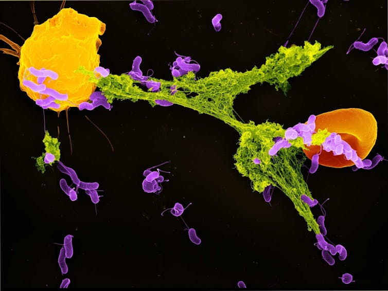
Once S. aureus, which is usually an asymptomatic colonizer, breaches our defenses, its various strategies of disrupting and evading the immune response become overwhelming, and it can spread to all tissue types in the body. The Monteith Lab examines how neutrophils detect these pathogens and modify their inflammatory response to combat their spread.
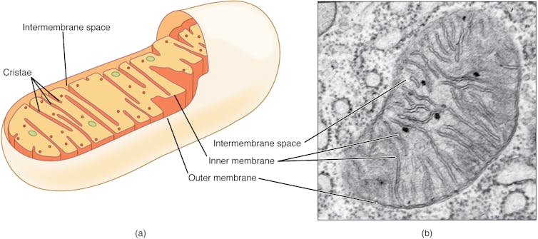
The lab also examines disruptions to mitochondrial homeostasis in immune cells related to SLE-mediated tissue damage. Understanding the molecular changes associated with disease could inform novel treatments for SLE, an autoimmune disease of which the cause is unknown.
The third realm of research in the lab involves attempting to characterize how changes in the metabolic controls of hematopoiesis are associated with myelodysplastic syndromes (MDS). MDS is associated with cytopenia, bone marrow failure, and even acute myeloid leukemia.
“There’s this very complex system of cells that evolve to work together, that are there to cause inflammation as well as resolve it,” Monteith said. “So, you’ve got this balance that occurs, but if things get out of balance, that’s when disease happens. Figuring out the factors driving imbalance is what drives me as a scientist.”
This interdisciplinary approach to studying disease at the microscopic level came as a result of Monteith’s path through research. His interest in both biophysics and immunology started in his undergraduate studies at Bradley University. During his PhD and post-doctoral research at the University of North Carolina at Chapel Hill, he worked on studying autoimmune disease, which informs his current work on SLE. His further post-doctoral study of S. aureus at Vanderbilt University also contributed to the questions his lab seeks to answer now.
“My favorite part as a scientist through the years, as a trainee from grad school to post-doc, is making that discovery that someone else hasn’t seen before. Being the first person to see that is what drove me, but that’s changed a little bit since being a faculty,” he said. Now a big part of what drives him is when members of his lab show him the new phenotypes and data they’ve found, being a part of their joy and helping them strategize their next direction.
Monteith’s broad interest across various aspects of immunology is making microbiology research more exciting than ever.
Investing in our Future
Dear Friends, Alumni, and Current Members of the University of Tennessee, Knoxville Department of Microbiology:
Cellular life, from microbes to man, is endlessly fascinating and a resource for meeting the challenges of our future. In this edition of our newsletter, you will see some highlights about our faculty and graduate student researchers who are gaining vital knowledge by investigating both fundamental and applied questions. Some of this knowledge will have immediate benefits for society. However, the tremendous long-term return on investment in scientific discovery is what I find most thrilling about the research mission. It can take decades for a seemingly esoteric discovery to transform our world, and we must trust our investments in the people who are making those discoveries.
One of my favorite examples of the delay between fundamental discoveries and our full realization of their implications is the molecular biology revolution. Cloning, the ability to cut and splice DNA at specific sites, is based on research on the fate of DNA of bacterial viruses (phages) within infected bacterial cells. In the 1950s, pioneers—including Grete Kellenberger-Gujer, Werner Arber (Nobel, 1978), and Daisy Dussoix—discovered that incoming phage DNA could be restricted from entry by nucleases that cut DNA at specific sites (Citi and Berg, 2016 Bacteriophage 6:e1148805; Loenen et al. 2014 Nucleic Acids Res. 42:3). These “restriction enzymes” are the foundation of recombinant DNA technology, used, among other things, to create the bacterial expression strains that remain our major source of therapeutic insulin.
Other examples of how researchers’ curiosity led to major societal transformations include the discovery of Thermus aquaticus in Yellowstone National Park that led to PCR-based technologies, such as forensic DNA analyses that can identify missing persons or criminals. Similarly, the discovery that some bacterial genomes have clustered, regularly interspersed short palindromic repeats, ultimately known as CRISPRs, led to a powerful gene editing technology that is being used to treat cancer and sickle cell anemia.
A common theme among these examples is that they each start with teams of researchers working together to apply their deep background knowledge, curiosity, creativity, and hard work to answering questions at the edge of science. Another theme is that the discoveries required connecting the living world around us to the laboratory—
with natural isolates of organisms, collected from the environment, being brought into the lab and studied to understand what is happening inside them. I am proud to say that these themes are in abundance in our department, and it is exciting to ponder the as-yet-unimaginable transformations that will be spurred by the work we are doing now. On behalf of the entire department, I offer thanks to those who invest in us for the future.
In this edition of our newsletter, you will hear the perspectives of faculty and graduate students on what it takes to succeed in research, and why we are driven to answer the big questions in science. You will learn about Assistant Professor Andrew Monteith’s investigations into the metabolic underpinnings of immunity in response to cancers, autoimmunity, and infectious diseases, and Professor Erik Zinser’s research on the physiology of and interactions among marine microbes and how technological advances are transforming the way we approach answering biological questions in these vast ecosystems. You also will learn from graduate student Mikayla Mangrum about her work on understanding the cell surfaces of the pathogenic fungus Candida albicans, toward identifying new treatments, and from recent PhD graduate Liz Glasgo, who developed new genetics tools that facilitated her investigations on the physiology of the marine bacterium Vibrio natriegens. Graduate student Toyosi Nimat Ajide-Bamigboye has focused her attention on a critical issue facing the globe: the impacts of microplastics on the microbiology and health of soil ecosystems.
This year we bid fond farewell to Distinguished Lecturer Elizabeth McPherson, who will be retiring after 30 years of dedicated service to our department. She has taught tens of thousands of undergraduate students, mentored hundreds of graduate students, and helped many new instructors at all levels find their footing in the classroom. She developed and continues to update and publish laboratory protocol books. Over the course of her career, she has received numerous Outstanding Undergraduate Teaching Awards from the department. Her contributions to our department are deep and broad, and we will miss her tremendously. Best wishes on a happy retirement Elizabeth!
I am pleased to announce the arrival in fall 2024 of three new assistant professors, Carolyn Ibberson, Sara Clasen, and Zach Burcham. Each brings new research and instructional expertise in the microbiology of human and other ecosystems. We also welcome Haley Dylewski (PhD ’24), who will join our instructional team. We look forward to sharing with you their exciting progress and news in future newsletters!
Heidi Goodrich-Blair
David and Sandra White Professor and
Head of Microbiology
$1.82 million NIH Grant Funding Lindsey Burcham’s Women’s Health Research
by Amy Beth Miller
With a $1.82 million National Institutes of Health grant, Assistant Professor Lindsey Burcham is leading interdisciplinary research on the vaginal microbiome, which can affect maternal, fetal, and pediatric health.
“We are incorporating techniques in microbial genetics/molecular biology, analytical chemistry, and in vitro and in vivo models to learn more about how microbes persist in the vaginal mucosa,” Burcham explained.
“This work will allow us to learn more about the availability of micronutrients in the vaginal tract and understand how vaginal microbes sense nutrient availability, respond to nutrient fluctuations, and share these nutrients within the community,” she said.
The five-year Maximizing Investigators’ Research Award (MIRA) began in 2023 and will run until June 2028.
Cultures and Computer Models
Burcham also is using computer simulations, or in silico models, in collaboration with UT microbiology Assistant Professor Zach Burcham to predict metabolite exchange between vaginal microbes.
“These models allow us to develop testable hypotheses to understand more about how microbes may interact with each other and behave in the host,” Lindsey Burcham said.
The researchers also will use synthetic communities, or controlled co-cultures of microbes, to study microbial interactions and to understand how individual microbes may impact the overall function of the microbial community.
Foundations for Exploration
The MIRA funding provides flexibility for investigators to be creative and work toward big-picture questions, Lindsey Burcham said. “The work outlined here will set the foundation for understanding more about the vaginal environment and microbe-microbe interactions, but I envision this is a starting point. We have already started some exciting new projects investigating other environmental factors within the vaginal tract, and we will go where the data take us.”
She has helped to assemble a collaborative “bench-to-bedside” research team including Zach Burcham and physicians Kim Fortner, Callie Reeder, and Logan Riley, as well as Associate Professor Jill Maples from the University of Tennessee Medical Center’s Department of Obstetrics and Gynecology.
Lindsey Burcham has been curious about microbes and motivated to understand how they work since she was an undergraduate student, and her personal life influenced her research interests.
“My own pregnancies fueled my curiosity for understanding the vaginal tract and the impact of microbes on vaginal health,” she said. “Now as the leader of a research team, I am excited to be able to use my lab and resources to promote women’s health research and to normalize the discussion around vaginal health.”
Research and Mentorship: Frank May Supported as an Emerging Scholar
by Randall Brown
Microbiology PhD student Frank May earned support as an SEC Emerging Scholar from the University of Tennessee Graduate School for the 2024–2025 academic year. This program provides $25,000 in fellowship support plus professional development, networking, and mentoring for chosen students in their final year of doctoral work.
As one of the three UT PhD students supported by this program for the year, May will also receive travel support to attend professional conferences.
“I would like to congratulate Frank May on this important award, which sends a positive message about microbiology and the natural sciences at UT,” said Professor Kate Jones, dean, Division of Natural Sciences and Mathematics in the College of Arts and Sciences. “His excellence in research, teaching, and outreach positions him to be a future leader in his chosen field.”
The financial support of this fellowship will give May more opportunity to focus on research as he completes his PhD program.
“This will enable me to spend the majority of my time working in the lab, both performing my own experiments and mentoring undergraduate students in conducting theirs,” said May.
Professor Heidi Goodrich-Blair, head of the Department of Microbiology, praises May’s dedication to scientific discovery and discourse and to student classroom and research success.
“He is a talented researcher who is untangling the complex relationships among bacterial viruses and plasmids and the cells they infect, and the higher order impact (e.g., on ecosystems) of these relationships,” said Goodrich-Blair. “His findings will have far-reaching ramifications because such impacts are ubiquitous among all cellular life on Earth.”
May’s research seeks to understand how mobile bacterial DNA alters the ability of bacteriophage—viruses that infect bacteria—to infect their hosts.
“The model organism we use is from the marine environment, giving us a better understanding of the impact of these mobile pieces of DNA, called plasmids, on this environment,” said May. “Additionally, these findings can also be extrapolated further to help us identify and understand bacterial defense strategies to viral infection.”
Goodrich-Blair also notes May’s mentorship of numerous undergraduates in independent research and his enthusiastic engagement in academic and community outreach and in organizing and participating in departmental activities.
“It cannot be understated how influential May has been on departmental committees and in representing his fellow graduate students on important issues,” she said.
May will use the support to pair outreach and professional development to represent UT microbiology at academic conferences—and return with fresh perspectives for the Vols he mentors.
“This award will assist me in attending the American Society for Microbiology Conference for Undergraduate Educators and the Annual Biomedical Research Conference for Minoritized Students paired conferences,” he said. “These meetings highlight new and evolving practices that better engage undergraduate students in an inclusive and equitable manner. This opportunity will expose me to new strategies that will help me grow as a biology educator to better serve future students.”
Steven Wilhelm & Brittany Zepernick Published in ‘The Conversation’
Losing winter ice is changing the Great Lakes food web – here’s how light is shaping life underwater
Winters on the Great Lakes are harsh – so much so that the scientists who work there often focus on the summer months, when tiny microbes at the base of the food chain were thought to be most productive.
However, emerging research is changing our understanding of these winter ecosystems and shining a light on a vibrant world of winter activity just below the ice.
Scientists discovered in the early 2000s that communities of diatoms – tiny photosynthesizing algae – were thriving in the light under the wind-swept lake ice. But, it turns out, that was only part of the story.
As the Great Lakes’ winter ice disappears – it hit record lows in the winter of 2023-24 – new analyses show that some diatoms appear to have a different way to create energy and survive in the dark, turbid ice-free water until summer.
These microbes are crucial to the Great Lakes’ health. They clean the water of pollutants and are the first step in the complex food web that supports a fishery that powers part of a regional economy. Changes here can have widespread effects on the lakes’ ecology and direct economic effects on surrounding communities.
Oozing up from the ice
Interest in life under the ice erupted in 2007, when an international team of scientists onboard a Canadian Coast Guard icebreaker noticed something unusual as the ship worked its way through the Lake Erie ice.
As the ice broke, dark brown water oozed up from the lake. It was teeming with diatoms.
There had been sporadic studies of the winter microbes in the past, but limnologists – scientists who study lakes – didn’t have the tools to fully understand the microbes’ behavior until recently.
For the past five years, the Joint Genome Institute of the U.S. Department of Energy has supported a molecular biology project that sequenced the RNA of all the microorganisms from samples collected from Lake Erie to address how these organisms survived winter months and might adapt, or not, to future climate scenarios. New observations about how diatoms may be using light are now emerging from this effort.
Using proteins common in animals’ eyes
Normally we think of diatoms as organisms that use sunlight to convert carbon dioxide into living material by photosynthesis. They’re pervasive in summer across the Great Lakes, where they help feed the lakes’ multibillion-dollar sport and commercial fisheries.
During winter, diatoms can create energy from the light that filters through the wind-swept ice. However, when the ice is not present in winter, the diatoms are mixed into lake water that can, at times, be best described as chocolate milk. Light penetrates poorly through this murky water, and the diatoms get less of the specific wavelengths of light that drive photosynthesis.
We collected samples in the winter of 2019-2020 to compare how diatom communities in open waters differed from those that live under ice. We were surprised that when ice wasn’t present, some diatoms were using a different form of energy acquisition – driven by a pigment called rhodopsin.
Rhodopsins are light-responsive proteins that are perhaps best known as a key component of the eyes of animals. In marine systems, it was shown in 2001 that these proteins are involved in generating energy in bacterial cells, specifically producing adenosine triphosphate, or ATP. ATP is a chemical that organisms use as a power supply for many cellular processes, leading to its nickname as the “molecular currency” of living cells.
It now seems that some Lake Erie diatoms use this energy generation mechanism to augment light-limited photosynthesis in ice-free winter months.
Differences in the two processes may be important: Photosynthesis helps cells fix carbon to produce new biomass as well as cellular energy in the form of ATP. With rhodopsins, while ATP is produced, there is no direct carbon fixation.
This means that cells can likely persist but not grow in these murky waters. But in biology, survival is everything: If an organism’s competitors do not survive harsh conditions but the organism does, there will be more nutrients when conditions improve. To this end, the rhodopsins in these diatoms appear to be as much a survival mechanism and an opportunity to persist in murky, ice-free winter conditions.
Watching lake life evolve as the climate changes
As we move into a warmer climate and ice-free era for Lake Erie and other Northern temperate lakes, this data suggests that, over time, the diatoms that thrived in ice-covered lakes may be replaced by diatoms with rhodopsins in winter months.
The consequences of this change are potentially manifold: Small changes at the base of the food web can affect fisheries. Moreover, some diatoms are known to produce compounds that are toxic to wildlife and humans.
We have only guesses at this point as to how changes in algal species will alter fisheries, tourism and coastal resource management in the long run. How algal communities change over time is a response to many drivers, and light is just one. But having the chance to watch this change from the beginning creates a unique opportunity to understand the effect of a warming climate on the Great Lakes and similar lakes around the world.
Steven Wilhelm, Professor of Microbiology, University of Tennessee; Brittany Zepernick, Postdoctoral Researcher in Microbiology, University of Tennessee, and Robert Michael McKay, Director and Professor, Great Lakes Institute for Environmental Research, University of Windsor
This article is republished from The Conversation under a Creative Commons license. Read the original article.